Anatomy Of Chest And Stomach - Anatomy : The embryologic and anatomic basis of modern surgery.. The stomach is a muscular, hollow organ in the gastrointestinal tract of humans and many other animals, including several invertebrates. Gross anatomy the stomach is a rounded, hollow organ located just inferior to the diaphragm in the left part of the abdominal cavity. What happens to fats, carbs & protein in foods. Stomach, saclike expansion of the digestive system, between the esophagus and the small intestine; This page provides an overview of the chest muscle group.
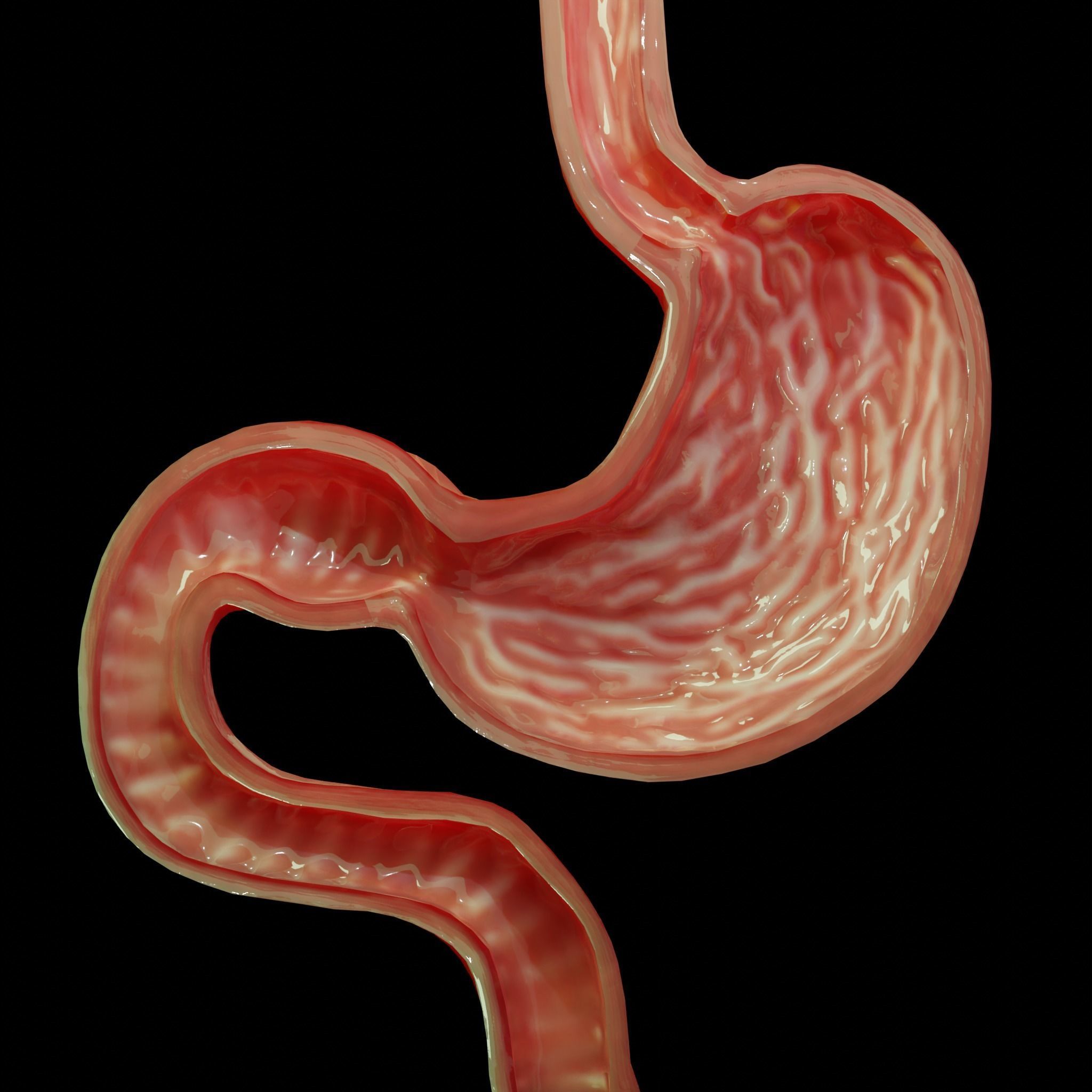
In the anatomy of stomach, it is the uppermost portion, forming the upper curvature of the organ. This type of ct scan uses a lower radiation level than a conventional. It can help you understand our world more detailed and specific. Its size and shape changes from time to time depending on the volume of its contents (food/fluid). False flatter chest fluctuating fluoride.
Interactive Thorax and Abdomen Human Anatomy in 3D - YouTube from i.ytimg.com Decker ga, plessis d du. The stomach consists of several important anatomical parts. Layers serosa or visceral peritoneum: Learn more from webmd about the anatomy of the stomach, along with illnesses that affect the stomach and tests to diagnose stomach problems. It is an internal organ between the esophagus and the small intestines. Swensen fund for innovation in teaching. It can help you understand our world more detailed and specific. If we were to locate it on our the principal function of this sphincter is to prevent food and stomach acids from regurgitating up the these include a burning sensation in the chest (heartburn), piercing or diffuse abdominal pain.
Three layers outer longitudinal middle circular inner oblique submucosa mucosa.
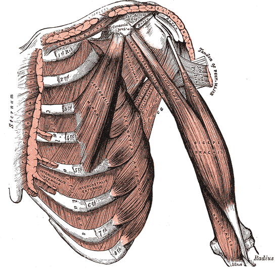
Lee mcgregor's synopsis of surgcial anatomy. The stomach lies within the superior aspect of the abdomen. Ingested food enters the stomach from the esophagus via the cardiac orifice, falling into gastric juice produced by the stomach. Stomach blood supply arterial blood supply: The stomach is part of the digestive system. It is an internal organ between the esophagus and the small intestines. Note that the stomach bubble is under the left hemidiaphragm. To duodenum regions cardiac fundus body pyloric. The stomach consists of several important anatomical parts. Muscles of shoulder anatomy spinous process of c7 vertebra, levator scapulae muscle, rhomboid minor i wanted to focus on the structure of the muscle on the chest and stomach. Anatomy of the stomach, gallbladder, and pancreas. The stomach is a muscular organ located on the left side of the upper abdomen. Protective framework for parts of the chest involved with brea… chest or thorax.
There are two sphincters of the stomach, located at each orifice. Decker ga, plessis d du. Its size and shape changes from time to time depending on the volume of its contents (food/fluid). Learning anatomy isn't easy but it will vastly improve your ability to draw. Muscles of shoulder anatomy spinous process of c7 vertebra, levator scapulae muscle, rhomboid minor i wanted to focus on the structure of the muscle on the chest and stomach.
Trunk Muscles | Boundless Anatomy and Physiology from s3-us-west-2.amazonaws.com The embryologic and anatomic basis of modern surgery. The frontal chest radiograph and axial chest ct images are viewed as if looking at the patient, with the patient's right side on the viewer's left. The upper portion of the trunk between the neck and the abdomen. In the anatomy of stomach, it is the uppermost portion, forming the upper curvature of the organ. What happens to fats, carbs & protein in foods. Three layers outer longitudinal middle circular inner oblique submucosa mucosa. Stimulated by the acidic condition in the stomach and certain other factors, these cells release pepsin enzyme in its inactive form, called pepsinogen, which then carries out the digestion of proteins. It can help you understand our world more detailed and specific.
Swensen fund for innovation in teaching.
Layers serosa or visceral peritoneum: Protective framework for parts of the chest involved with brea… chest or thorax. This type of ct scan uses a lower radiation level than a conventional. Grant jcb, basmajian jv, slonecker ce. The stomach lies within the superior aspect of the abdomen. What are parts of the stomach & the stomach anatomy. The stomach receives food from the esophagus. There are two sphincters of the stomach, located at each orifice. Anatomynote.com found chest muscle anatomy from plenty of anatomical pictures on the internet. Swensen fund for innovation in teaching. The wall of the stomach is structurally similar to other parts of the digestive tube, with the exception that the stomach has an extra oblique layer of smooth muscle inside the circular layer, which aids in the. Anatomical structures are visible as. What happens to fats, carbs & protein in foods.
We hope you will use this picture in the study and. To duodenum regions cardiac fundus body pyloric. It is an internal organ between the esophagus and the small intestines. What are parts of the stomach & the stomach anatomy. Anatomy of the stomach, gallbladder, and pancreas.
3D model Stomach cross sectional anatomy | CGTrader from img2.cgtrader.com Its size and shape changes from time to time depending on the volume of its contents (food/fluid). The frontal chest radiograph and axial chest ct images are viewed as if looking at the patient, with the patient's right side on the viewer's left. There are two sphincters of the stomach, located at each orifice. Layers serosa or visceral peritoneum: What happens to fats, carbs & protein in foods. The main function of the stomach involves mechanical and chemical digestion of ingested food. The pancreas stores and secretes pancreatic juice into the duodenum to complete the chemical digestion of food that began in the mouth and stomach. We hope you will use this picture in the study and.
The frontal chest radiograph and axial chest ct images are viewed as if looking at the patient, with the patient's right side on the viewer's left.
This page provides an overview of the chest muscle group. To duodenum regions cardiac fundus body pyloric. Protective framework for parts of the chest involved with brea… chest or thorax. This type of ct scan uses a lower radiation level than a conventional. The stomach is the third stage in the digestive process. The main function of the stomach involves mechanical and chemical digestion of ingested food. The stomach is part of the digestive system. What happens to fats, carbs & protein in foods. Nervous tissue in the submucosa monitors the contents of the stomach and controls smooth muscle contraction and secretion of digestive substances. The embryologic and anatomic basis of modern surgery. We think this is the most useful anatomy picture anatomy is the amazing science. Layers serosa or visceral peritoneum: Swensen fund for innovation in teaching.
It is located in the anterior portion of the abdominal cavity in most the stomach serves as a temporary receptacle for the storage and mechanical distribution of food before it is passed into the intestine anatomy of chest. The wall of the stomach is structurally similar to other parts of the digestive tube, with the exception that the stomach has an extra oblique layer of smooth muscle inside the circular layer, which aids in the.


0 Komentar